

Life under the microscope: Nikon photo contest shows creepy closeups, stunning imagery
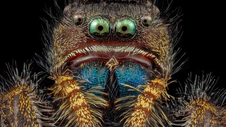
Bold jumping spider (Phidippus audax) by Dr. Andrew Posselt, University of California San Francisco (Nikon Small World)
The winners of the 48th annual Nikon Small World contest for photography under the microscope are in, and one astonishing "image of distinction" is raising hairs around the world.
The contest, founded in 1974 to recognize excellence in microphotography, received nearly 1,300 entries from 72 countries. The submissions are judged on originality, informational content, technical proficiency and visual impact.
"Each year, Nikon Small World receives an array of microscopic images that exhibit exemplary scientific technique and artistry. This year was no exception," said Eric Flem, communications and CRM manager for Nikon Instruments. "At the intersection of art and science, this year’s competition highlights stunning imagery from scientists, artists, and photomicrographers of all experience levels and backgrounds from across the globe."
The embryonic, fluorescent hand of a Madagascar Giant Day Gecko, taken by Grigorii Timin and Dr. Michel Milinkovitch at the University of Geneva, Switzerland, took home first place.
"A visually stunning and painstaking technique, Timin used image stitching to merge hundreds of images together to create the final image of his gecko," judges said in a news release. "The final result gives a glimpse into the hidden beauty and complexity of the gecko, highlighting the nerves in a cyan color and the bones, tendons, ligaments, skin and blood cells in a range of warmer colors."
RELATED: There are more ants on Earth than the combined total of birds, mammals, study finds
Dr. Eugenijus Kavaliauskas’s extreme close-up of an ant, one of dozens of photos honored as an "image of distinction," is staggering. The image was created in Lithuania and used a reflective light technique to show the ant’s eyes, hair and antennas.
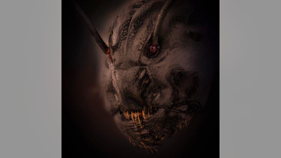
Extreme closeup of an ant by Dr. Eugenijus Kavaliauskas, Lithuania
Second place went to Dr. Caleb Dawson for his image of breast tissue showing contractile myoepithelial cells wrapped around milk-producing alveoli.
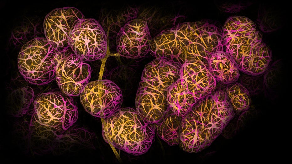
Second Place: Breast tissue showing contractile myoepithelial cells wrapped around milk-producing alveoli by Dr. Caleb Dawson, WEHI, The Walter and Eliza Hall Institute of Medical Research, Australia (Nikon Small World)
Satu Paavonsalo and Dr. Sinem Karama from the University of Helsinki in Finland won third place for their image of blood vessel networks in the intestine of an adult mouse.
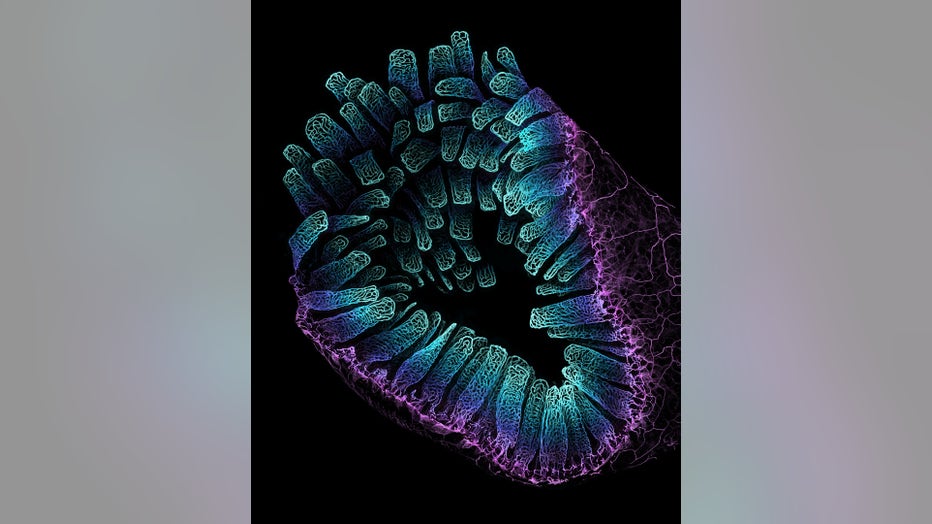
Third Place: Blood vessel networks in the intestine of an adult mouse by Satu Paavonsalo & Dr. Sinem Karaman, University of Helsinki (Nikon Small World)
"The Nikon Small World Competition is a great opportunity to share how impressive nature is on a microscopic level, not only within a scientific community but also with the general public," Timin said.
Click here for more Nikon Small World 2022 photo winners.
In 2011, Nikon Small World launched a sister competition — Nikon Small World in Motion — to showcase technology advances for recording movies or digital time-lapse photography through the microscope. You can see those winners here.